|
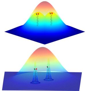
The fluorescent signals from two nearby objects are superimposed by diffraction and imaged as a single feature. The ability to image individual probes separately means that their positions can be determined much more accurately to reconstruct the whole structure.
[Credit: University of Heidelberg] |
Fluorescence microscopy is a widely used method for studying cellular structures. However, the so-called diffraction limit prevents detailed examination of these structures. Objects separated by less than 0.3 µm from one another cannot be resolved. Recently, different methods have been reported to overcome this limit, among them stochastic optical reconstruction microscopy (STORM). Here the cell structures are labelled with fluorescent dyes that respond with fluorescence emission upon stimulation by light of a certain wavelength. High resolution of approx. 0.02 µm can be achieved by switching off the majority of the dyes and leaving only a small number of them on. In this way, the light emitted by neighbouring dyes is no longer superimposed. This dye-switching is also controlled by light. The dyes left on can now be individually resolved and their positions can be determined by mathematical analysis to a very high accuracy of approx. 0.003 µm. Multiple repetition of this procedure supplies precise information about the location of all the dyes, thus permitting high-resolution reconstruction of the cell structures under investigation.
This method puts several demands on the microscope and the light sources used. Switching the respective dyes requires either different laser lines (excitation wavelength) or high light intensities or both. This can lead to problems in the investigation of living cells. Accordingly, the team headed by Heidelberg chemist Dr. Dirk-Peter Herten has replaced the switching of dyes via laser light with a light-independent process. To this end, the scientists adjusted a chemical probe for the detection of copper ions in such a way that the probe and its fluorescent properties can be used to mark cellular structures. If copper(II) binds to the probe, its fluorescence is suppressed. This binding of the copper(II) ion is reversible, thus restoring the fluorescence of the probe. The microscopic investigation of cell structures is therefore controlled by a reversible chemical reaction.
The scientists have called this method CHIRON – chemically improved resolution for optical nanoscopy. According to Dr. Herten, it means that microscopy methods like STORM can be simplified to such an extent that additional laser lines and high light intensities become superfluous. All that is required is for the probe to be located in a cellular environment to which minimal amounts of copper sulphate can be added, e.g. fixed cells. “This opens up new applications for high-resolution microscopy that were formerly impracticable due to technical restrictions,” says Dr. Herten. “Our probes can be used on many microscopes.”